Mesothelioma Cat Cytology - Pdf Mesothelioma In Domestic Animals Cytological And Anatomopathological Aspects : mesothelioma is often diagnosed as a lung cancer or initially as pneumonia because several of the symptoms of mesothelioma are also symptoms of pneumonia and similar conditions.
Mesothelioma Cat Cytology - Pdf Mesothelioma In Domestic Animals Cytological And Anatomopathological Aspects : mesothelioma is often diagnosed as a lung cancer or initially as pneumonia because several of the symptoms of mesothelioma are also symptoms of pneumonia and similar conditions.. Diagnostic imaging is therefore of paramount importance in this. The dog presented a severe abdominal distension. This type is the type that occurs most often. Positron emission tomography (pet) scan. Cowell r l, tyler r d &
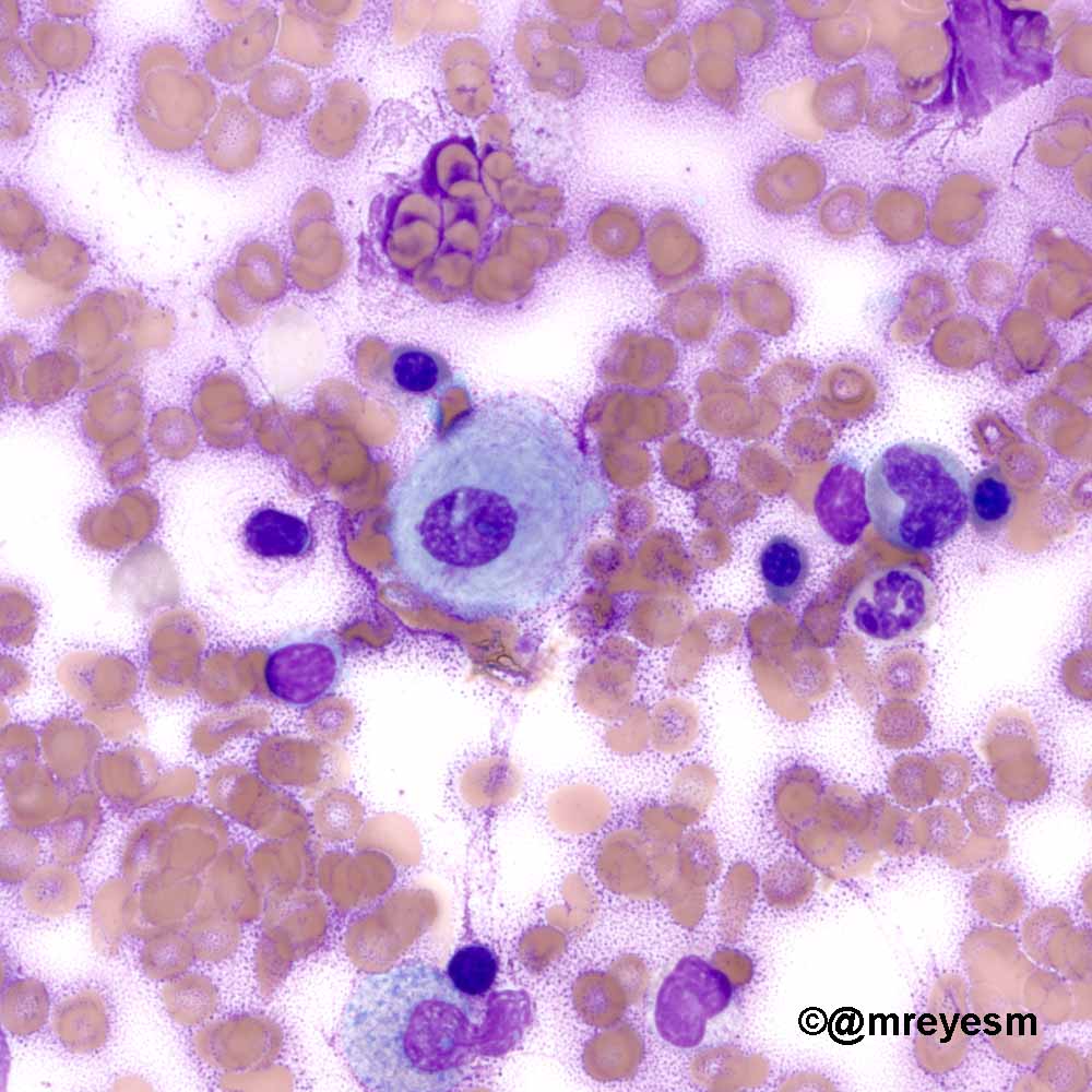
If a large amount of fluid is present, abnormal cells may be detected by cytology if this fluid is aspirated with a syringe. 1,2,3,4 this case showed the difficulties in the diagnosis of mesothelioma in domestic animals and how cytology sometimes has to be supported by other findings (clinical history, histology and immunohistochemistry) to achieve a correct diagnosis. (b) bronchoalveolar lavage specimen with strongyloides in an immunocompromised patient (papanicolaou stain, ×600). A ct (or cat) scan or an mri is usually performed. Chylous effusions are the result of leakage of lymph into the body cavity and may involve either the pleural or peritoneal space.
Pdf Mesothelioma In Domestic Animals Cytological And Anatomopathological Aspects from www.researchgate.net cytology, histopathology, and immunohistochemical analysis. This cancer is a form of mesothelioma that develops in the abdomen, specifically on the peritoneum (the lining of the abdominal cavity). In 2021 alone, it is estimated that 119,100 men and 116,660 women will be diagnosed with lung cancer, and 69,410 men and 62,470 women will die of this disease. A cat scan or an mri and cytology is then done to determine if a large amount of fluid and or abnormal cells are present. It usually begins to form in the cells in the glands, which are found in the lungs. For pleural fluid this is done by a pleural tap or chest drain, in ascites with an paracentesis or ascitic drain and in a pericardial effusion with A ct (or cat) scan or an mri is usually performed. Mesomark® was the first approved in 2007, making it the first blood test to be used for mesothelioma diagnosis.
Pleural effusion showing a cluster of neoplastic cells from a case of malignant mesothelioma.(papanicolaou x100 oil immersion) ovarian cancer cells in abdominal effusion.
A ct (or cat) scan or an mri is usually performed. All types require a mesothelioma biopsy for a definitive diagnosis. Pleural effusion showing a cluster of neoplastic cells from a case of malignant mesothelioma.(papanicolaou x100 oil immersion) ovarian cancer cells in abdominal effusion. A cat scan or an mri and cytology is then done to determine if a large amount of fluid and or abnormal cells are present. A ct (or cat) scan or an mri is usually performed. Cytological examination of the peritoneal fluid revealed anaplastic epithelioid cells. Isolated and pseudopapillary groups of neoplastic cells associated to psammoma bodies and inflammatory cells. Diagnostic testing for asbestos cancer. Immunohistochemical staining for pancytokeratin was positive for both internalized and host cells, Lung cancer is the third most common form of noncutaneous cancer in the united states and is the leading cause of cancer death in men and in women. Neoplastic conditions such as lymphoma, carcinoma, mesothelioma, and Diagnosis of thoracic lesions on the basis of history and physical examination is often challenging. Clinical findings and survival in 17 dogs studied retrospectively r.
Pleural fluid cytology is insensitive for the diagnosis of mesothelioma, although it may reveal other malignant cell types. mesothelioma is often diagnosed as a lung cancer or initially as pneumonia because several of the symptoms of mesothelioma are also symptoms of pneumonia and similar conditions. If a large amount of fluid is present, abnormal cells may be detected by cytology if this fluid is aspirated with a syringe. After mesothelioma has been diagnosed, tests are done to find out if cancer cells have spread to other parts of the body. mesothelioma is cancer that affects mesothelioma, a tissue that connects the different organs of the body.
Eosin E Hemat Sohaibsyed8 Twitter from pbs.twimg.com If a large amount of fluid is present, abnormal cells may be detected by cytology if this fluid is aspirated with a syringe. Chylous effusions are the result of leakage of lymph into the body cavity and may involve either the pleural or peritoneal space. As mesothelioma has a tendency to invade needle tracts, definitive diagnostic techniques (e.g. Diagnostic imaging is therefore of paramount importance in this. If a large amount of fluid is present, abnormal cells may be detected by cytology if this fluid is aspirated with a syringe. 1,2,3,4 this case showed the difficulties in the diagnosis of mesothelioma in domestic animals and how cytology sometimes has to be supported by other findings (clinical history, histology and immunohistochemistry) to achieve a correct diagnosis. This cancer is a form of mesothelioma that develops in the abdomen, specifically on the peritoneum (the lining of the abdominal cavity). Cell internalization was observed by cytology in two of these cases (the feline mammary tumour and the pleural effusion in the canine mesothelioma) and by histopathology in all but the canine mesothelioma.
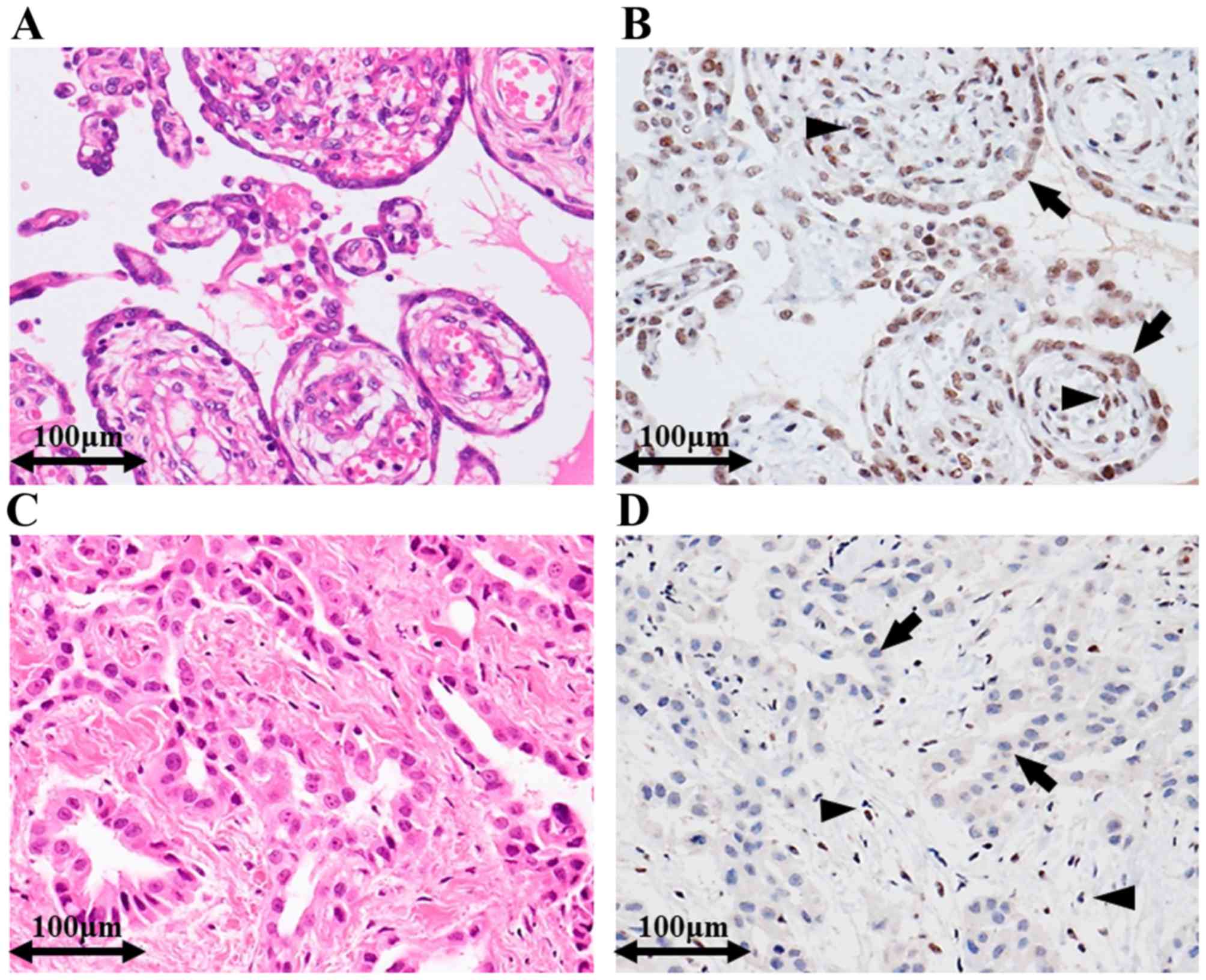
Malignant mesothelioma is an uncommon tumor arising mostly from the serosal surface of the pleural or peritoneal cavity .in most human patients, pleural mesothelioma is a locally advanced tumor and the invasion of the thoracic wall with rib involvement has been reported 2, 3.a locally invasive nature of malignant mesotheliomas has rarely been reported in dogs, while involvement of ribs has.
A ct (or cat) scan or an mri is usually performed. Already with cytology and the postdrainage imaging findings were consistent with mesothelioma. There is contralateral tracheal and mediastinal shift (red arrows). It is extremely rare, accounting for only about 20 percent of all mesothelioma cases annually, or about 660 cases per year in the united states. Mesothelial hyperplasia is often accompanied by pleural fibrosis (figure 23.24) in cases of subpleural inflammation and especially when inflammation is chronic active, and may occur as micropapillary fronds of mature connective tissue covered by a single layer of flattened to cuboidal mesothelial cells (figure 23.25). A ct (or cat) scan or an mri is usually performed. Tional mesothelioma interest group staging system of pleural mesothelioma, this condition, with or without rib destruction is described in stages iii and iv . It usually begins to form in the cells in the glands, which are found in the lungs. Diagnosing malignant mesothelioma can be difficult. Lung cancer is the third most common form of noncutaneous cancer in the united states and is the leading cause of cancer death in men and in women. A cat scan or an mri and cytology is then done to determine if a large amount of fluid and or abnormal cells are present. Pleural mesothelioma ( pleural mesothelioma ), which is a cancer that attacks the lining of the mesothelium of the lungs (pleura). Mesomark® was the first approved in 2007, making it the first blood test to be used for mesothelioma diagnosis.
The signs and symptoms depend upon the location of the tumor. Ghisleni g, roccabianca p, ceruti r, et al: Tional mesothelioma interest group staging system of pleural mesothelioma, this condition, with or without rib destruction is described in stages iii and iv . Meinkoth j h (1999) diagnostic cytology and hematology of the dog and cat. 2 mesothelioma is a neoplastic proliferation of the serosal cell layer lining the peritoneal, pericardial, or pleural spaces.
Utility Of Survivin Bap1 And Ki 67 Immunohistochemistry In Distinguishing Epithelioid Mesothelioma From Reactive Mesothelial Hyperplasia from www.spandidos-publications.com Isolated and pseudopapillary groups of neoplastic cells associated to psammoma bodies and inflammatory cells. If you suspect you have this cancer, you will almost certainly undergo one of them: Malignant mesothelioma is an uncommon tumor arising mostly from the serosal surface of the pleural or peritoneal cavity .in most human patients, pleural mesothelioma is a locally advanced tumor and the invasion of the thoracic wall with rib involvement has been reported 2, 3.a locally invasive nature of malignant mesotheliomas has rarely been reported in dogs, while involvement of ribs has. This cancer is a form of mesothelioma that develops in the abdomen, specifically on the peritoneum (the lining of the abdominal cavity). Positron emission tomography (pet) scan. Factors involved in diagnosing mesothelioma include: Immunohistochemical staining for pancytokeratin was positive for both internalized and host cells, Background malignant mesothelioma is an uncommon tumor arising mostly from the serosal surface of the pleural or peritoneal cavity .in most human patients, pleural mesothelioma is a locally advanced tumor and the invasion of the thoracic wall with rib involvement has been reported 2, 3.a locally invasive nature of malignant mesotheliomas has rarely been reported in dogs, while involvement.
Cowell r l, tyler r d &
mesothelioma is often diagnosed as a lung cancer or initially as pneumonia because several of the symptoms of mesothelioma are also symptoms of pneumonia and similar conditions. mesothelioma and adenocarcinoma are both forms of cancer but are significantly different diseases. Immunohistochemical staining for pancytokeratin was positive for both internalized and host cells, Four cases of tumors in which cell internalization was frequently visualized are reported: 1,2,3,4 this case showed the difficulties in the diagnosis of mesothelioma in domestic animals and how cytology sometimes has to be supported by other findings (clinical history, histology and immunohistochemistry) to achieve a correct diagnosis. In cats, mesothelioma have been only rarely described. A ct (or cat) scan or an mri is usually performed. A biopsy is the only definitive way to confirm a mesothelioma diagnosis. If a large amount of fluid is present, abnormal cells may be detected by cytology if this fluid is aspirated with a syringe. mesothelioma testing commonly includes imaging scans, biopsies and blood tests. Cytological examination of the peritoneal fluid revealed anaplastic epithelioid cells. Pleural mesothelioma ( pleural mesothelioma ), which is a cancer that attacks the lining of the mesothelium of the lungs (pleura). (b) bronchoalveolar lavage specimen with strongyloides in an immunocompromised patient (papanicolaou stain, ×600).

0 Comments